
Spinal Stenosis: Understanding Neurogenic Claudication and Treatment Options
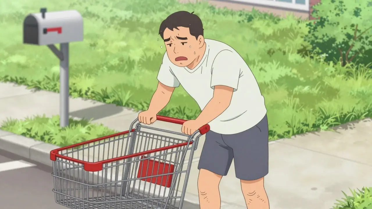
Walking to the mailbox used to be a simple errand. Now, you find yourself stopping every few steps, leaning forward, or waiting for that heavy, cramping feeling in your legs to fade. If this sounds familiar, you might be dealing with neurogenic claudication, a hallmark symptom of lumbar spinal stenosis. It is not just back pain; it is a specific pattern of leg discomfort triggered by posture and movement. Understanding the difference between this condition and vascular issues is the first step toward finding relief.
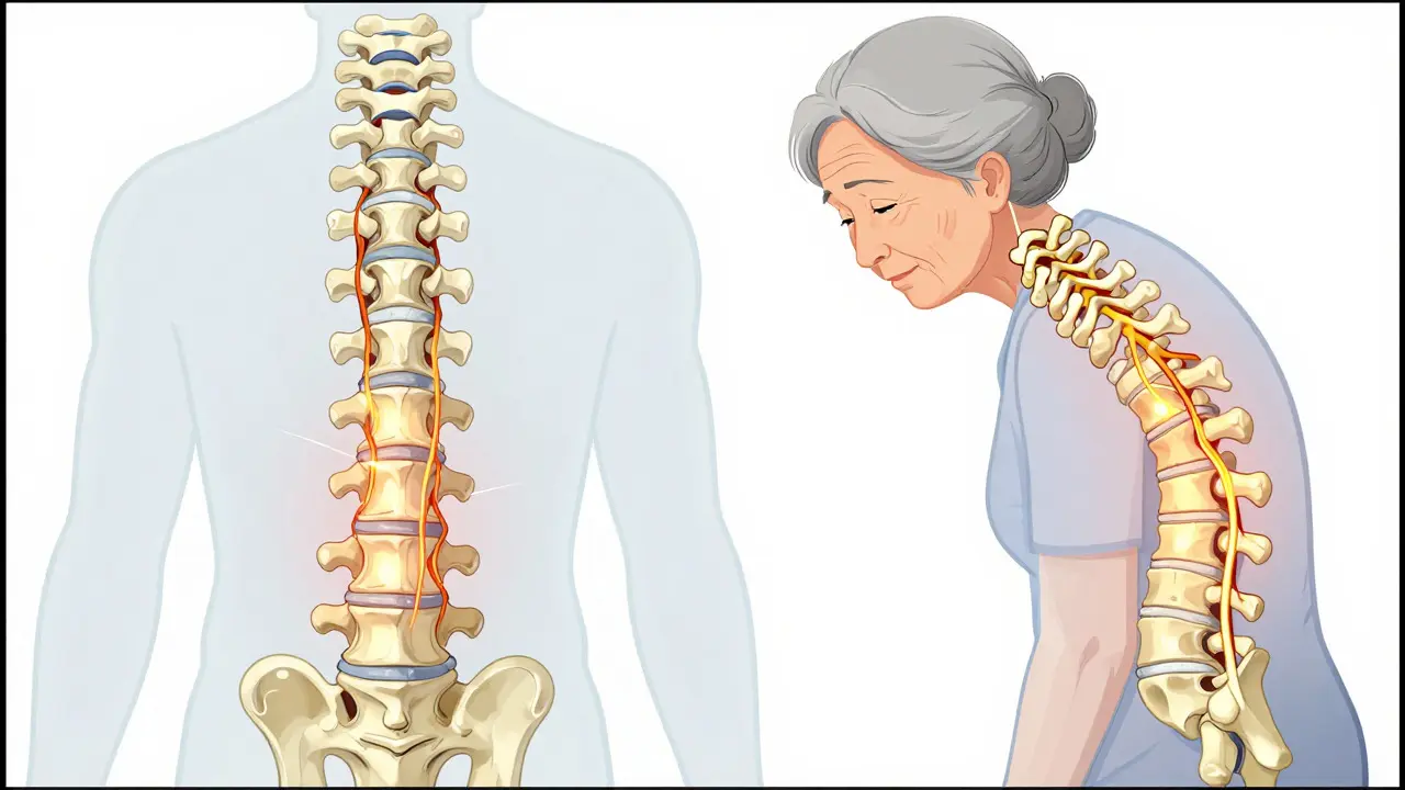
The core problem here is mechanical compression. As we age, the spinal canal can narrow due to degenerative changes like bone spurs or thickened ligaments. This narrowing squeezes the nerve roots in the lower back. When you stand upright or walk, the space tightens further, cutting off blood flow to the nerves or causing direct pressure. The result is pain, numbness, or weakness in the buttocks and legs. But unlike a heart attack or blocked artery, the fix isn't about clearing a vessel; it's about changing the geometry of your spine to give those nerves room to breathe.
Key Takeaways
- Neurogenic claudication is defined by leg pain that worsens with walking or standing but improves significantly when sitting or bending forward.
- The "shopping cart sign"-relief from leaning over a cart-is a classic diagnostic clue present in up to 85% of cases.
- Accurate diagnosis is critical because treating it as a vascular issue leads to ineffective care.
- Conservative management, including flexion-based physical therapy, resolves symptoms for many patients within 3-6 months.
- Surgery, such as laminectomy, is considered when conservative options fail, offering significant improvement for 70-80% of selected patients.
Recognizing the Symptoms: More Than Just Leg Pain
Many people assume any leg pain while walking means they have bad arteries. However, neurogenic claudication has a distinct signature. The pain typically starts in the buttocks or thighs and radiates down the legs. It feels like heaviness, cramping, tingling, or weakness. The key differentiator is positional relief. If you stop walking and simply stand still, the pain often persists. But if you sit down or bend forward at the waist (the "simian stance"), the symptoms subside rapidly.
This forward-flexion relief happens because bending opens up the spinal canal, reducing pressure on the compressed nerves. You might notice you walk better pushing a shopping cart or a walker. This is known as the "shopping cart sign." In clinical settings, this behavior is so common that it serves as a strong indicator of lumbar stenosis rather than peripheral artery disease. If your foot pulses are strong and symmetric, but you need to hunch over to walk comfortably, the issue is likely neural, not vascular.
Physical examination usually reveals normal reflexes or mild lower motor neuron signs, such as diminished ankle jerks. A straight leg raise test is typically negative, which helps rule out a herniated disc. Clinicians also look for wasting in the small muscles of the foot, specifically the extensor digitorum brevis. Visible wasting here is a reliable bedside marker for underlying severe stenosis, signaling that the nerves have been under compression long enough to cause muscle atrophy.
Diagnosis: Why Imaging Alone Isn't Enough
Getting a diagnosis often involves a mix of history-taking and imaging. Doctors will ask specific questions: Does the pain improve when you sit? Do you feel better riding a bicycle? These details help distinguish neurogenic causes from vascular ones. While an MRI is the gold standard for visualizing the spinal canal, it has limitations. Studies show that up to 67% of asymptomatic individuals have abnormal MRI findings consistent with stenosis. This means having a narrowed canal on an image doesn't automatically mean it's the source of your pain.
Therefore, the correlation between imaging and symptoms is crucial. A doctor looks for a match between what the MRI shows and what you report. For example, if the MRI shows central canal stenosis at L4-L5 and you have bilateral leg pain relieved by flexion, the diagnosis is solid. If the imaging shows severe stenosis but your symptoms don't fit the pattern, other causes must be investigated. This multi-faceted approach ensures that treatment targets the actual problem, avoiding unnecessary interventions.
| Feature | Neurogenic Claudication (Spinal) | Vascular Claudication (Arterial) |
|---|---|---|
| Pain Trigger | Walking, standing upright | Walking, exertion |
| Relief Mechanism | Sitting, bending forward | Stopping activity, any rest position |
| Foot Pulses | Normal, symmetric | Diminished or absent |
| Posture Impact | Forward flexion improves symptoms | Posture does not significantly change symptoms |
| Underlying Cause | Nerve compression in spinal canal | Blood flow restriction in arteries |
Conservative Management: The First Line of Defense
For most patients, surgery isn't the first step. Conservative care forms the backbone of initial treatment, aiming to manage symptoms and maintain function without invasive procedures. This phase typically lasts 3 to 6 months. The goal is to reduce inflammation, strengthen supporting muscles, and modify daily activities to minimize nerve compression.
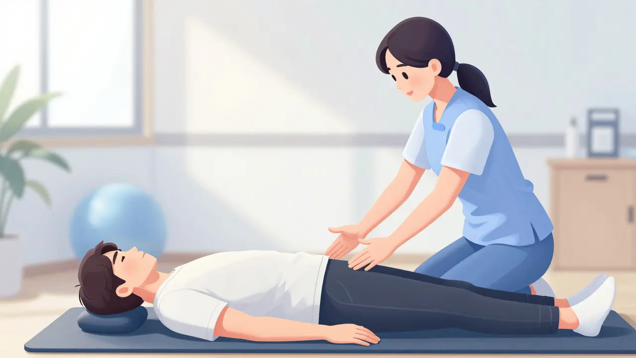
Physical therapy is central to this approach. Unlike general fitness routines, therapy for spinal stenosis focuses on flexion-based exercises. Movements that open the front of the spine and close the back, such as pelvic tilts or gentle knee-to-chest stretches, help relieve pressure. Patients are often taught posture training to avoid prolonged extension, which narrows the canal. Adherence to these programs is vital; inconsistent participation often leads to perceived failure of conservative care. Many patients find that simply adjusting how they stand and move throughout the day provides significant relief.
Pain medication may also play a role. Non-steroidal anti-inflammatory drugs (NSAIDs) can help with associated inflammation, though their effect on neural pain varies. In some cases, neuropathic agents like gabapentin or pregabalin are prescribed to calm irritated nerves. Epidural steroid injections offer another option for temporary relief. By injecting corticosteroids around the affected nerve roots, clinicians can reduce local inflammation and swelling. Success rates for temporary pain relief range from 50% to 70%, providing a window of opportunity for patients to engage more effectively in physical therapy.
Surgical Interventions: When to Consider Decompression
If conservative measures fail after several months, or if neurological deficits progress, surgery becomes a viable option. The primary goal of surgical intervention is decompression-creating more space for the nerves. The most common procedure is a laminectomy, where part of the lamina (the bony arch at the back of the vertebra) is removed. This directly widens the spinal canal, relieving pressure on the neural elements.
Minimally invasive techniques have gained traction in recent years. Procedures using smaller incisions and specialized instruments aim to reduce tissue trauma and speed up recovery. Data from the American Association of Neurological Surgeons indicates a 35% increase in minimally invasive decompression procedures between 2018 and 2022. These approaches often result in less postoperative pain and shorter hospital stays compared to traditional open surgery.
Outcomes are generally positive for appropriately selected patients. Studies suggest that 70% to 80% of individuals experience significant improvement in walking distance and pain levels after surgery. However, surgery is not a cure-all. It addresses the mechanical compression but does not reverse age-related degeneration. Recurrence of symptoms is possible, especially if adjacent levels develop stenosis later. Therefore, patient selection is critical. Surgery is best suited for those with clear anatomical stenosis matching their symptoms and who have exhausted conservative options.
Living with Spinal Stenosis: Practical Strategies
Managing lumbar spinal stenosis is a lifestyle adjustment as much as a medical one. Understanding your triggers allows you to navigate daily activities more comfortably. Walking remains beneficial, but pacing is key. Instead of long, uninterrupted walks, try shorter intervals with breaks where you sit or lean forward. This keeps you active without provoking severe symptoms.
Your environment matters too. If you work at a desk, ensure your chair supports a slight forward tilt. Standing desks should allow you to shift weight and avoid rigid upright postures. When doing household chores, use tools that keep your hands elevated, mimicking the forward-flexion posture that relieves symptoms. Bicycles, particularly recumbent ones, are excellent alternatives for cardiovascular exercise because the seated, leaned-forward position naturally opens the spinal canal.
Maintaining a healthy weight also reduces stress on the spine. Excess body mass increases the load on the lumbar vertebrae, potentially worsening stenosis symptoms. Combining weight management with targeted core strengthening helps stabilize the spine, reducing the dynamic compression that occurs during movement. Remember, the goal is not to eliminate all movement but to optimize how you move.
Frequently Asked Questions
How do I know if my leg pain is from spinal stenosis or bad arteries?
The key difference is relief mechanism. If bending forward or sitting makes the pain go away quickly, it is likely neurogenic (spinal). If stopping walking and standing still provides relief regardless of posture, it is more likely vascular (arterial). Checking foot pulses is also helpful; strong pulses suggest a spinal cause.
Is surgery dangerous for older adults with spinal stenosis?
Surgery carries risks, but modern minimally invasive techniques have reduced complications. For older adults, the decision depends on overall health and symptom severity. Many patients over 70 undergo successful decompression surgeries with good outcomes, provided they are medically cleared for anesthesia.
Can spinal stenosis get worse if I don't treat it?
Yes, degenerative changes tend to progress slowly over time. Without management, the narrowing can worsen, leading to increased frequency of symptoms and potential permanent nerve damage if compression becomes severe. Early intervention with conservative care can slow progression and preserve function.
What is the "shopping cart sign"?
It refers to the phenomenon where patients with spinal stenosis can walk longer distances if they lean over a shopping cart or walker. This forward-flexion posture opens the spinal canal, relieving pressure on the nerves and allowing for more comfortable ambulation.
How long does it take to see results from physical therapy?
Most patients begin to notice improvements in flexibility and pain management within 4 to 6 weeks of consistent therapy. Full assessment of response typically takes 3 to 6 months. Consistency is crucial; missing sessions can delay progress and make it harder to determine if the therapy is working.
Emmanuel Umana
August 27, 2026 AT 23:21The geometry of the spine dictates the flow of life. When the canal narrows, the spirit is trapped in the flesh. We must respect the body's architecture.
Lilian Binda
August 29, 2026 AT 01:19why do americans always think they need surgery for everything its just a back pain you old fools get some rest and stop complaining about your knees and backs
Amy Zalkin
August 29, 2026 AT 21:04oh my gosh this hit home so hard i was doing the shopping cart thing at the grocery store and felt like a total weirdo but turns out it was just my spine being dramatic. love how bending forward helps it feels like cheating the system a bit lol
Alex Brown
August 31, 2026 AT 18:00It is fascinating how posture alters neural compression. The distinction between vascular and neurogenic claudication is not merely academic; it is the difference between treating a symptom and addressing the root cause. We should value the diagnostic precision described here.
Kaylyn Mello
September 2, 2026 AT 07:35i totally agree with the point about physical therapy being key. i started doing those knee-to-chest stretches and honestly it made a huge difference within a few weeks. consistency is really everything though if you skip days it gets worse again
joyce Hogewoning
September 3, 2026 AT 22:17so basically if you cant walk without hunching over like a goblin in a dungeon you probably have spinal stenosis right? because that is exactly what my mom does and she refuses to see a doctor saying it is just age but i am telling her it might be something more serious than just getting old and creaky like a rusty gate
Thhomas Fox
September 5, 2026 AT 12:22Great breakdown. The 'shopping cart sign' is a brilliant diagnostic clue. I've seen many patients who were misdiagnosed as vascular when it was actually neural. Flexion-based PT is a game changer for them.
Owen John
September 7, 2026 AT 03:42Of course, the MRI is a tool of the state, designed to pathologize normal aging. But let us not forget that the true cure lies in rejecting the modern upright posture imposed by industrial capitalism. Only by returning to the primal crouch can we liberate our nerves from the tyranny of verticality. It is all connected, isn't it?
Maneesh kv
September 7, 2026 AT 04:31this is so true 😂 i had this issue last year and thought i had bad arteries but turned out to be stenosis. the forward bend trick saved me from unnecessary tests. keep moving friends 💪🔥
John Park
September 8, 2026 AT 04:14let's be real here the whole concept of 'conservative management' is just a way for insurance companies to delay the inevitable cash grab of surgery. why wait 6 months when you could just cut the bone now and get back to work faster? efficiency is king and patience is for the weak.
Kathleen McGrath
September 9, 2026 AT 21:45I found this article really helpful! I have been struggling with leg pain when I walk for months. It is great to know that sitting or bending forward can help. I am going to try the recumbent bike idea next week. Thanks for sharing!
Lemuel Gomez
September 10, 2026 AT 21:00One small note, however... while the MRI correlation is important, don't we also need to consider genetic factors in ligament thickening? I have read that certain populations are predisposed to faster degenerative changes. It adds another layer to the diagnosis process, doesn't it?
Fabian Saldana
September 11, 2026 AT 21:49An excellent overview of a complex condition. Understanding the mechanical nature of neurogenic claudication empowers patients to take control of their recovery. Let us continue to advocate for accurate diagnoses and evidence-based care.
Colin Finch
September 13, 2026 AT 20:58This is such a relief to read! I was terrified it was my heart or legs failing. Turns out it was just my spine being grumpy. The tip about standing desks is gold - I will definitely adjust mine to allow for that slight forward tilt. Thanks for the clear explanation!